Bestand:Paracentrotus lividus myogenesis.jpg

Grootte van deze voorvertoning: 538 × 600 pixels. Andere resoluties: 215 × 240 pixels | 430 × 480 pixels | 689 × 768 pixels | 1.138 × 1.269 pixels.
Oorspronkelijk bestand (1.138 × 1.269 pixels, bestandsgrootte: 1,72 MB, MIME-type: image/jpeg)
| Dit is een bestand van Wikimedia Commons. Onderstaande beschrijving komt van de beschrijving van het bestand daar. |
Beschrijving
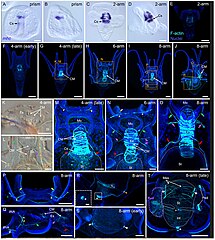
| Beschrijving | FIGURE 10. Myogenesis in Paracentrotus lividus during the embryonic and larval periods. Developmental stages are as follows: (A,B) prism stage (prism); (C–E) 2-arm pluteus stage (2-arm); (F,G,K,M) 4-arm pluteus stage (4-arm); (H,L,N) 6-arm pluteus stage (6-arm); (I,J,O–T) 8-arm pluteus stage (8-arm). The use of (early) or (late) associated with the stage names simply highlights here more specific periods during the 4- or 8-arm pluteus stages. In (A–D), images were acquired using bright-field, differential interference contrast light, and they correspond to embryos and larvae labeled for the muscle terminal differentiation gene myosin heavy chain (mhc). In (E–J,M–T), images are maximum intensity projections of confocal z-stacks of larvae co-labeled for F-actin (muscles; cyan) and DNA (nuclei; blue), and they correspond to projections of the entire specimen. In (K,L), images were acquired using bright-field light microscopy. In (A), the embryo is in ventral view, with the animal pole up. In (B,D,J,K,Q,R), embryos and larvae are in left view, with either the animal pole up and the ventral side left in (B) or with the anterior pole up and the ventral side left in (D,K,Q,R) or with the anterior pole right and the ventral side up in (J). In (C,E–I,L–P,S,T), larvae are in anterior view, with the ventral side up. (K–T) Close-ups of the regions outlined by orange boxes in (G–J). (K–O,Q) Close-ups of the esophageal region. (P) Close-up of the oral hood. (R) Close-up of the intestinal region. ((R) inset) Close-up of the anal sphincter. (S,T) Close-ups of the stomach region. In (K–O,T), the white dotted line outlines the larval digestive tract. In (L,N,O,Q), green arrows highlight the posterior dilator muscles, and purple arrows mark the lateral muscles. In (M–O,Q), red arrows indicate the ventrolateral processes (or longitudinal musculature). In (M,N,P,Q), yellow arrowheads mark the anterior dilator muscles (or star-shaped muscles). In (N,P,Q), yellow double arrowheads highlight the preoral dilator muscles. In (R,T), orange arrowheads mark the pyloric sphincter, and the white asterisk marks the anal sphincter. In (S,T), white arrows indicate the lateral stomach muscles. In (T), the purple dotted line delineates the adult rudiment. In (H,J,M,N,P,Q,S,T), the F-actin staining detected along the ciliary band and the epaulettes corresponds to a counterstain of the apex of the cuboidal cells and thus not to muscle cells. Scale bar: (A–D,M–O) 30 μm; (E,F,K,L,P–T) 50 μm; (G–J) 100 μm; ((R) inset) 10 µm. CM: circumesophageal muscle; Cs: cardiac sphincter; Es: esophagus; Int: intestine; lAlA: left anterolateral arm; lPrA: left preoral arm; Mes: mesentery; Mo: mouth; Ped: pedicellariae; Rud: adult rudiment; St: stomach. |
| Datum | |
| Bron |
https://www.frontiersin.org/articles/10.3389/fcell.2022.966408/full Developmental atlas of the indirect-developing sea urchin Paracentrotus lividus: From fertilization to juvenile stages, Front. Cell Dev. Biol., 31 October 2022 Sec. Morphogenesis and Patterning Volume 10 - 2022, https://doi.org/10.3389/fcell.2022.966408 |
| Auteur | Laurent Formery, Axel Wakefield, Maeva Gesson, Ludovic Toisoul, Guy Lhomond, Laurent Gilletta, Régis Lasbleiz, Michael Schubert, Jenifer C. Croce1 |
Licentie
Dit bestand is gelicenseerd onder de Creative Commons Naamsvermelding 4.0 Internationaal licentie.
- De gebruiker mag:
- Delen – het werk kopiëren, verspreiden en doorgeven
- Remixen – afgeleide werken maken
- Onder de volgende voorwaarden:
- naamsvermelding – U moet op een gepaste manier aan naamsvermelding doen, een link naar de licentie geven, en aangeven of er wijzigingen in het werk zijn aangebracht. U mag dit op elke redelijke manier doen, maar niet zodanig dat de indruk wordt gewekt dat de licentiegever instemt met uw werk of uw gebruik van zijn werk.

|
Dit bestand, dat oorspronkelijk toegevoegd was op een externe website, is nog niet beoordeeld door een moderator of reviewer om te bevestigen dat de opgegeven licentie geldig is. Zie Category:License review needed voor meer instructies.
|
Bestandsgeschiedenis
Klik op een datum/tijd om het bestand te zien zoals het destijds was.
| Datum/tijd | Miniatuur | Afmetingen | Gebruiker | Opmerking | |
|---|---|---|---|---|---|
| huidige versie | 6 mrt 2024 12:58 | | 1.138 × 1.269 (1,72 MB) | Rasbak | {{Information |description=FIGURE 10. Myogenesis in Paracentrotus lividus during the embryonic and larval periods. Developmental stages are as follows: (A,B) prism stage (prism); (C–E) 2-arm pluteus stage (2-arm); (F,G,K,M) 4-arm pluteus stage (4-arm); (H,L,N) 6-arm pluteus stage (6-arm); (I,J,O–T) 8-arm pluteus stage (8-arm). The use of (early) or (late) associated with the stage names simply highlights here more specific periods during the 4- or 8-arm pluteus stages. In (A–D), images were a... |
Bestandsgebruik
Dit bestand wordt op de volgende pagina gebruikt: