Bestand:Node Cilia Are Posteriorly Tilted and Positioned primitive node.png

Grootte van deze voorvertoning: 383 × 599 pixels. Andere resoluties: 153 × 240 pixels | 307 × 480 pixels | 491 × 768 pixels | 655 × 1.024 pixels | 2.020 × 3.157 pixels.
Oorspronkelijk bestand (2.020 × 3.157 pixels, bestandsgrootte: 4,01 MB, MIME-type: image/png)
| Dit is een bestand van Wikimedia Commons. Onderstaande beschrijving komt van de beschrijving van het bestand daar. |
Beschrijving
| Beschrijving |
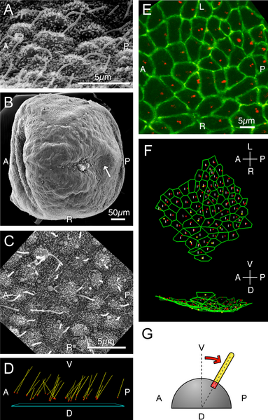
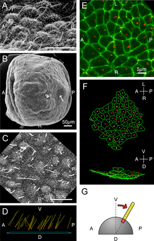
Figure 3. Node Cilia Are Posteriorly Tilted and Positioned (A) Scanning electron micrograph of the wild-type node. Note that cilia emanate from the posterior part of the cells. The view angle is about 30° with respect to the horizontal line. (B, C) Scanning electron micrograph of the iv/iv node. (C) is a high-magnification picture of the region in (B) indicated by an arrow. (D) Deduced tilt of iv/iv node cilia after stereography from multiple-tilt scanning electron micrograph images. Yellow lines indicate cilia, red dots their root positions, and a blue square a plane best-fit to the node surface. When we calculated the tilt of the cilia, we separated the tilt into A-P (anterior–posterior) and L-R components. The average tilt was 26.6° in A-P axis (toward the posterior) and 0.06° in L-R axis (towards the right). (E) Immunofluorecence image of node cells shown as projection of 3D confocal data stack. Basal bodies and cell boundaries are shown by immunofluorescence against γ-tubulin (red) and ZO-1 (green), respectively. (F) 3D reconstruction of (E) viewed from ventral side (top) and right side (bottom), showing posterior bias of basal body positions. White lines divide the cells into the anterior and the posterior halves. Basal bodies located in the anterior and the posterior are shown in yellow and red, respectively. (G) Speculative interpretation of posterior bias of basal bodies in orientation and position of the node cilia. Because the node cells are somewhat rounded, if basal bodies were located at the posterior part of these cells, it would result in posteriorly tilted cilia even though the basal bodies remain perpendicular to the plasma membrane |
| Datum | |
| Bron | https://doi.org/10.1371/journal.pbio.0030268 (2005) De Novo Formation of Left–Right Asymmetry by Posterior Tilt of Nodal Cilia. PLoS Biol 3(8): e268. |
| Auteur | Nonaka S, Yoshiba S, Watanabe D, Ikeuchi S, Goto T, Marshall WF, et al. |

|
Dit bestand, dat oorspronkelijk toegevoegd was op een externe website, is nog niet beoordeeld door een moderator of reviewer om te bevestigen dat de opgegeven licentie geldig is. Zie Category:License review needed voor meer instructies.
|
Copyright: © 2005 Nonaka et al. This is an open-access article distributed under the terms of the Creative Commons Attribution License, which permits unrestricted use, distribution, and reproduction in any medium, provided the original work is properly cited.
Licentie
Dit bestand is gelicenseerd onder de Creative Commons Naamsvermelding 4.0 Internationaal licentie.
- De gebruiker mag:
- Delen – het werk kopiëren, verspreiden en doorgeven
- Remixen – afgeleide werken maken
- Onder de volgende voorwaarden:
- naamsvermelding – U moet op een gepaste manier aan naamsvermelding doen, een link naar de licentie geven, en aangeven of er wijzigingen in het werk zijn aangebracht. U mag dit op elke redelijke manier doen, maar niet zodanig dat de indruk wordt gewekt dat de licentiegever instemt met uw werk of uw gebruik van zijn werk.
Bestandsgeschiedenis
Klik op een datum/tijd om het bestand te zien zoals het destijds was.
| Datum/tijd | Miniatuur | Afmetingen | Gebruiker | Opmerking | |
|---|---|---|---|---|---|
| huidige versie | 4 mei 2024 22:30 | | 2.020 × 3.157 (4,01 MB) | Rasbak | {{Information |description=Figure 3. Node Cilia Are Posteriorly Tilted and Positioned (A) Scanning electron micrograph of the wild-type node. Note that cilia emanate from the posterior part of the cells. The view angle is about 30° with respect to the horizontal line. (B, C) Scanning electron micrograph of the iv/iv node. (C) is a high-magnification picture of the region in (B) indicated by an arrow. (D) Deduced tilt of iv/iv node cilia after stereography from multiple-tilt scanning electron... |
Bestandsgebruik
Dit bestand wordt op de volgende pagina gebruikt: