Bestand:Free-living amebic infections.png
Oorspronkelijk bestand(1.365 × 1.170 pixels, bestandsgrootte: 715 kB, MIME-type: image/png)


Deze biologieafbeelding zou opnieuw moeten worden aangemaakt als een SVG-bestand door vectorafbeeldingen te gebruiken. Dit heeft een aantal voordelen; zie Commons:Media for cleanup voor meer informatie. Als er een SVG-formaat van deze afbeelding bestaat, dan deze graag uploaden. Nadat u dit heeft gedaan, gelieve dit sjabloon te vervangen door het sjabloon {{vector version available|nieuwe bestandsnaam.svg}} op deze afbeeldingspagina.

Beschrijving

Beschrijving
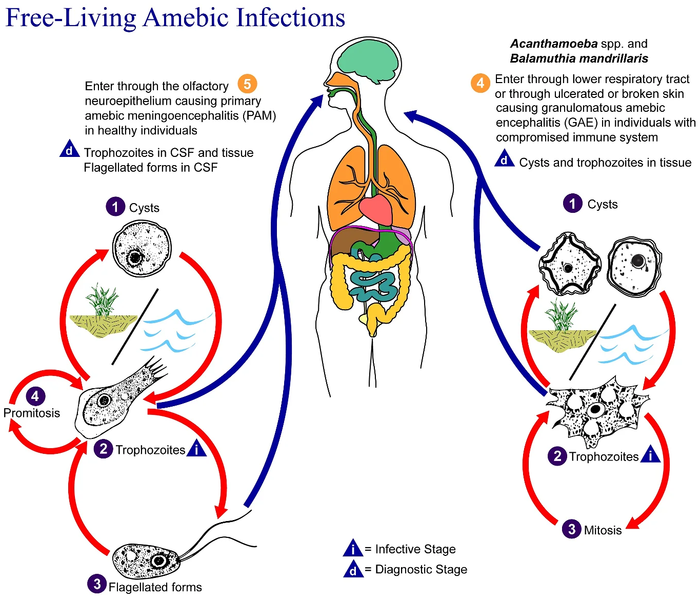
English: This is an illustration of the life cycle of the parasitic agents responsible for causing “free-living” amebic infections.

For a complete description of the life cycle of these parasites, select the link below the image or paste the following address in your address bar: http://www.dpd.cdc.gov/dpdx/HTML/FreeLivingAmebic.htm Free-living amebae belonging to the genera Acanthamoeba, Balamuthia, and Naegleria are important causes of disease in humans and animals. Naegleria fowleri produces an acute, and usually lethal, central nervous system (CNS) disease called primary amebic meningoencephalitis (PAM). N. fowleri has three stages, cysts (1) , trophozoites (2) , and flagellated forms (3) , in its life cycle. The trophozoites replicate by promitosis (nuclear membrane remains intact) (4) . Naegleria fowleri is found in fresh water, soil, thermal discharges of power plants, heated swimming pools, hydrotherapy and medicinal pools, aquariums, and sewage. Trophozoites can turn into temporary flagellated forms which usually revert back to the trophozoite stage. Trophozoites infect humans or animals by entering the olfactory neuroepithelium (5) and reaching the brain. N. fowleri trophozoites are found in cerebrospinal fluid (CSF) and tissue, while flagellated forms are found in CSF.

Acanthamoeba spp. and Balamuthia mandrillaris are opportunistic free-living amebae capable of causing granulomatous amebic encephalitis (GAE) in individuals with compromised immune systems. Acanthamoeba spp. have been found in soil; fresh, brackish, and sea water; sewage; swimming pools; contact lens equipment; medicinal pools; dental treatment units; dialysis machines; heating, ventilating, and air conditioning systems; mammalian cell cultures; vegetables; human nostrils and throats; and human and animal brain, skin, and lung tissues. B. mandrillaris however, has not been isolated from the environment but has been isolated from autopsy specimens of infected humans and animals. Unlike N. fowleri, Acanthamoeba and Balamuthia have only two stages, cysts (1) and trophozoites (2) , in their life cycle. No flagellated stage exists as part of the life cycle. The trophozoites replicate by mitosis (nuclear membrane does not remain intact) (3) . The trophozoites are the infective forms and are believed to gain entry into the body through the lower respiratory tract, ulcerated or broken skin and invade the central nervous system by hematogenous dissemination (4). Acanthamoeba spp. and Balamuthia mandrillaris cysts and trophozoites are found in tissue.
Datum
Bron

Deze media is afkomstig van de Centra voor ziektebestrijding en -preventie's Beeldbank volksgezondheid (PHIL), met identificatienummer '#3412.

NB: Niet alle PHIL-afbeeldingen zijn publiek domein; Zorg ervoor dat u de auteursrechtstatus controleert en auteurs en inhoudsproviders vermeldt.


العربية | Deutsch | English | македонски | slovenščina | +/−

Auteur
  • Photo Credit:
  • Content Providers(s): CDC/Alexander J. da Silva, PhD/Melanie Moser
Toestemming
(Hergebruik van dit bestand)
English: None - This image is in the public domain and thus free of any copyright restrictions. As a matter of courtesy we request that the content provider be credited and notified in any public or private usage of this image.
Andere versies

Licentie

Public domain
Deze afbeelding is een werk van de Centers for Disease Control and Prevention, onderdeel van de United States Department of Health and Human Services, genomen of gemaakt tijdens de officiële werkzaamheden van een werknemer. Als werk van de Federale overheid van de Verenigde Staten, bevindt de afbeelding zich in het publiek domein.

eesti  Deutsch  čeština  español  português  English  français  Nederlands  polski  slovenščina  suomi  македонски  українська  日本語  中文(简体)‎  中文(繁體)‎  العربية  +/−

Oorspronkelijk uploadlogboek

Verplaatst vanaf en.wikipedia naar Commons door Optigan13.

De oorspronkelijke beschrijving van deze afbeelding stond hier. Alle volgende gebruikersnamen verwijzen naar en.wikipedia.
  • 2006-05-04 01:09 Keenan Pepper 518×435×8 (31658 bytes) Free-living_amebic_infections.gif, converted to [[PNG]] by [[netpbm]].

Bijschriften

Beschrijf in één regel wat dit bestand voorstelt

Items getoond in dit bestand

beeldt af

image/png

Bestandsgeschiedenis

Klik op een datum/tijd om het bestand te zien zoals het destijds was.

Datum/tijdMiniatuurAfmetingenGebruikerOpmerking
huidige versie2 feb 2023 11:24Miniatuurafbeelding voor de versie van 2 feb 2023 11:241.365 × 1.170 (715 kB)Materialscientisthttps://answersingenesis.org/biology/microbiology/the-genesis-of-brain-eating-amoeba/
20 jul 2008 08:30Miniatuurafbeelding voor de versie van 20 jul 2008 08:30518 × 435 (31 kB)Optigan13{{Information |Description={{en|This is an illustration of the life cycle of the parasitic agents responsible for causing “free-living” amebic infections. For a complete description of the life cycle of these parasites, select the link below the image

Geen enkele pagina gebruikt dit bestand.

Globaal bestandsgebruik

De volgende andere wiki's gebruiken dit bestand:

Metadata