Bestand:Diagrams and images of human embryos at the gastrula stage.png

Grootte van deze voorvertoning: 587 × 599 pixels. Andere resoluties: 235 × 240 pixels | 470 × 480 pixels | 752 × 768 pixels | 1.003 × 1.024 pixels | 2.006 × 2.048 pixels | 3.128 × 3.193 pixels.
Oorspronkelijk bestand (3.128 × 3.193 pixels, bestandsgrootte: 804 kB, MIME-type: image/png)
| Dit is een bestand van Wikimedia Commons. Onderstaande beschrijving komt van de beschrijving van het bestand daar. |
Beschrijving
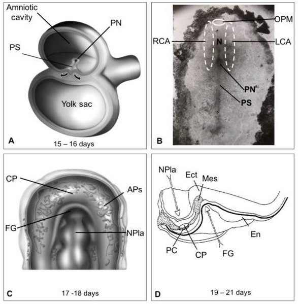
| Beschrijving | Figure 2. Diagrams and images of embryos at the gastrula stage. (A) Development of the trilaminar embryonic disk of chicken due to the migration (arrows) of epiblast cells through the primitive streak (PS). (B) A 16 ± 1-day embryo exhibiting the right (RCA) and left (LCA) cardiogenic areas spanning one-third of the primitive streak, the primitive node (PN), and the notochord (N) to the oropharyngeal membrane (OPM). (C) An 18 ± 1-day embryo showing the angiogenic plexuses (APs) arranged in the cardiac crescent, facing the neural plate (NPla). (D) Sagittal section of a 19–21-day human embryo with the first pair of somites showing the beginning of the development of the foregut (FG) and the neural plate. The somatopleure (SP), pericardial cavity (PC), and cardiogenic plate (CP) on the endoderm (En) of the yolk sac are also visible |
| Datum | |
| Bron | https://doi.org/10.3390/life13010165 Human Heart Morphogenesis: A New Vision Based on In Vivo Labeling and Cell Tracking. Life 2023, 13, 165. |
| Auteur | Villavicencio-Guzmán, L.; Sánchez-Gómez, C.; Jaime-Cruz, R.; Ramírez-Fuentes, T.C.; Patiño-Morales, C.C.; Salazar-García, M. |

|
Dit bestand, dat oorspronkelijk toegevoegd was op een externe website, is nog niet beoordeeld door een moderator of reviewer om te bevestigen dat de opgegeven licentie geldig is. Zie Category:License review needed voor meer instructies.
|
© 2023 by the authors. Licensee MDPI, Basel, Switzerland. This article is an open access article distributed under the terms and conditions of the Creative Commons Attribution (CC BY) license (https://creativecommons.org/licenses/by/4.0/).
Licentie
Dit bestand is gelicenseerd onder de Creative Commons Naamsvermelding 4.0 Internationaal licentie.
- De gebruiker mag:
- Delen – het werk kopiëren, verspreiden en doorgeven
- Remixen – afgeleide werken maken
- Onder de volgende voorwaarden:
- naamsvermelding – U moet op een gepaste manier aan naamsvermelding doen, een link naar de licentie geven, en aangeven of er wijzigingen in het werk zijn aangebracht. U mag dit op elke redelijke manier doen, maar niet zodanig dat de indruk wordt gewekt dat de licentiegever instemt met uw werk of uw gebruik van zijn werk.
Bestandsgeschiedenis
Klik op een datum/tijd om het bestand te zien zoals het destijds was.
| Datum/tijd | Miniatuur | Afmetingen | Gebruiker | Opmerking | |
|---|---|---|---|---|---|
| huidige versie | 5 mei 2024 02:22 | | 3.128 × 3.193 (804 kB) | Rasbak | {{Information |description= Figure 2. Diagrams and images of embryos at the gastrula stage. (A) Development of the trilaminar embryonic disk of chicken due to the migration (arrows) of epiblast cells through the primitive streak (PS). (B) A 16 ± 1-day embryo exhibiting the right (RCA) and left (LCA) cardiogenic areas spanning one-third of the primitive streak, the primitive node (PN), and the notochord (N) to the oropharyngeal membrane (OPM). (C) An 18 ± 1-day embryo showing the angiogenic pl... |
Bestandsgebruik
Dit bestand wordt op de volgende 2 pagina's gebruikt: