Bestand:Anaplastic astrocytoma.jpg
Grootte van deze voorvertoning: 800 × 552 pixels. Andere resoluties: 320 × 221 pixels | 640 × 442 pixels | 1.024 × 707 pixels | 1.200 × 828 pixels.
Oorspronkelijk bestand (1.200 × 828 pixels, bestandsgrootte: 200 kB, MIME-type: image/jpeg)
| Dit is een bestand van Wikimedia Commons. Onderstaande beschrijving komt van de beschrijving van het bestand daar. |
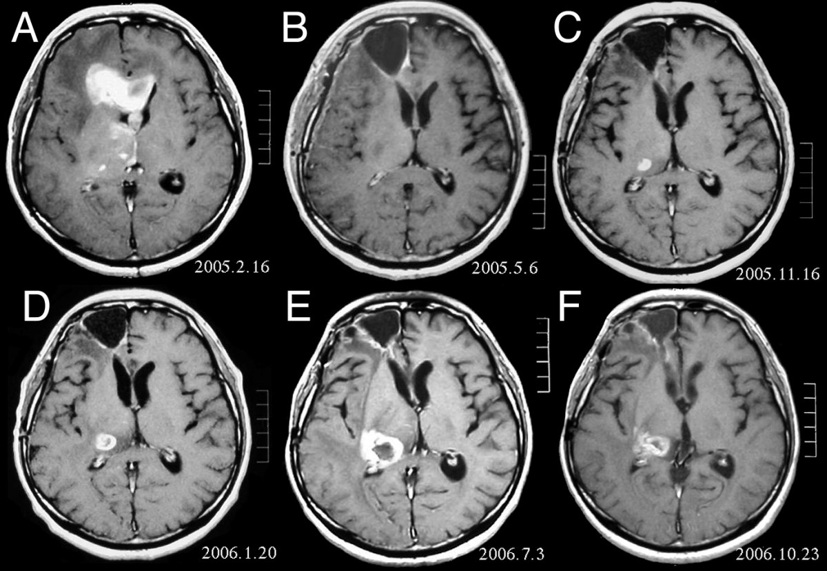
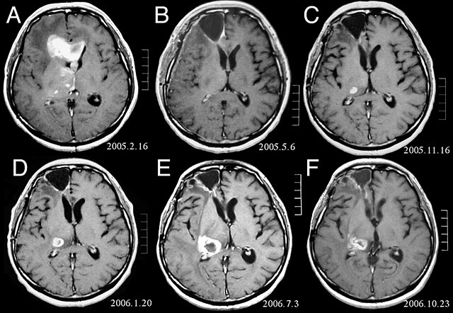
| Beschrijving | MRI of brain. (A) Initial MRI on February 16, 2005, shows a tumor in the right and left frontal lobe as well as the right thalamus. (B) MRI after surgery, radiation and chemotherapy. The tumor has completely disappeared except for slight enhancement adjacent to the surgical margin. (C) Recurrence of the thalamic tumor despite maintenance chemotherapy on November 16, 2005. (D) Increase in size of the thalamic tumor two months after stereotactic radiotherapy. (E) After 6 cycles of TMZ therapy, the thalamic lesion enlarged, and the patient developed dysarthria and hemiparesis. (F) After 2 courses of treatment with interferon-beta and TMZ, the tumor shows a partial response. |
| Datum | |
| Bron | Fujimaki T, Ishii H, Matsuno A, Arai H, Nakagomi T.Effectiveness of interferon-beta and temozolomide combination therapy against temozolomide-refractory recurrent anaplastic astrocytoma.World J Surg Oncol. 2007 Aug 4;5:89. PMID 17683572 doi:10.1186/1477-7819-5-89 |
| Auteur | Fujimaki T, Ishii H, Matsuno A, Arai H, Nakagomi T. |
| Toestemming (Hergebruik van dit bestand) |
BioMedCentral License |
Dit bestand is gelicenseerd onder de Creative Commons-licentie Naamsvermelding 2.0 Unported
- De gebruiker mag:
- Delen – het werk kopiëren, verspreiden en doorgeven
- Remixen – afgeleide werken maken
- Onder de volgende voorwaarden:
- naamsvermelding – U moet op een gepaste manier aan naamsvermelding doen, een link naar de licentie geven, en aangeven of er wijzigingen in het werk zijn aangebracht. U mag dit op elke redelijke manier doen, maar niet zodanig dat de indruk wordt gewekt dat de licentiegever instemt met uw werk of uw gebruik van zijn werk.
Bestandsgeschiedenis
Klik op een datum/tijd om het bestand te zien zoals het destijds was.
| Datum/tijd | Miniatuur | Afmetingen | Gebruiker | Opmerking | |
|---|---|---|---|---|---|
| huidige versie | 25 feb 2008 18:47 | | 1.200 × 828 (200 kB) | Filip em | {{Information |Description=MRI of brain. (A) Initial MRI on February 16, 2005, shows a tumor in the right and left frontal lobe as well as the right thalamus. (B) MRI after surgery, radiation and chemotherapy. The tumor has completely disappeared except f |
Bestandsgebruik
Dit bestand wordt op de volgende pagina gebruikt:
Globaal bestandsgebruik
De volgende andere wiki's gebruiken dit bestand:
- Gebruikt op ar.wikipedia.org
- Gebruikt op bg.wikipedia.org
- Gebruikt op cs.wikipedia.org
- Gebruikt op da.wikipedia.org
- Gebruikt op de.wikipedia.org
- Gebruikt op el.wikipedia.org
- Gebruikt op en.wikipedia.org
- Gebruikt op es.wikipedia.org
- Gebruikt op et.wikipedia.org
- Gebruikt op hi.wikipedia.org
- Gebruikt op hr.wikipedia.org
- Gebruikt op hu.wikipedia.org
- Gebruikt op it.wikipedia.org
- Gebruikt op kk.wikipedia.org
- Gebruikt op lb.wikipedia.org
- Gebruikt op lv.wikipedia.org
- Gebruikt op mk.wikipedia.org
- Gebruikt op mt.wikipedia.org
- Gebruikt op no.wikipedia.org
- Gebruikt op pl.wikipedia.org
- Gebruikt op ro.wikipedia.org
- Gebruikt op sk.wikipedia.org
- Gebruikt op sl.wikipedia.org
- Gebruikt op sq.wikipedia.org
- Gebruikt op sr.wikipedia.org